If the diagnosis was made by contrast enhanced MRI, then follow up is all you would do unless the arachnoid cyst becomes symptomatic (headaches, hydrocephalus or brain herniation as it getting bigger). If the diagnosis was made by CT or noncontrast enhanced MRI, then you need the MRI with contrast to exclude any enhancing solid component which would be diagnostic of pilocystic astrocytoma.
Don't be afraid of 破裂 -- which is exactly the surgical treatment method.
Hope that helps.
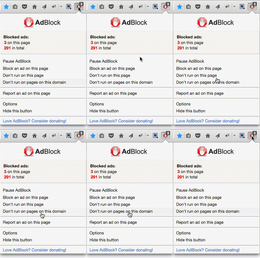
 选择“Disable on www.wenxuecity.com”
 选择“don't run on pages on this domain”

